

Best Place To Insert A Blood Clot Filter?
Inferior Vena Cava Filter Placement and Removal
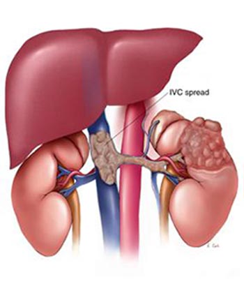
During Inferior Vena Cava (IVC) filter placement, a filtering device is placed within the IVC, a large vein in the abdomen that returns blood from the lower half of the torso to the heart. Blood clots in the veins of the legs and pelvis can occasionally travel to the lungs where they may crusade a pulmonary embolism or blockage. IVC filters aid reduce the risk of pulmonary embolism past trapping large clots and preventing them from reaching the middle and lungs. They are used in patients who don't reply to or cannot be given conventional medical therapy such as claret thinners.
Your doctor will instruct yous on how to prepare for the process. You will be advised on any changes to your regular medication schedule and whether you should not swallow or drink earlier your procedure. Tell your medico if there'southward a possibility you are significant and talk over any recent illnesses, medical atmospheric condition, allergies and medications you lot're taking. Get out jewelry at home and wear loose, comfortable wearable. You may be asked to wear a gown. Plan to take someone drive you dwelling house afterward.
- What is Inferior Vena Cava Filter Placement and Removal?
- What are some mutual uses of the procedure?
- How should I prepare?
- What does the equipment look like?
- How does the procedure work?
- How is the procedure performed?
- What will I experience during and afterward the process?
- Who interprets the results and how do I become them?
- What are the benefits vs. risks?
What is Inferior Vena Cava Filter Placement and Removal?
In an inferior vena cava filter placement procedure, interventional radiologists use image guidance to place a filter in the junior vena cava (IVC), the large vein in the abdomen that returns blood from the lower half of the trunk to the middle.
Claret clots that develop in the veins of the leg or pelvis, a status chosen deep vein thrombosis (DVT), occasionally suspension upwardly and large pieces of the clot tin can travel to the lungs. An IVC filter is a modest metal device that traps large clot fragments and prevents them from traveling through the vena cava vein to the heart and lungs, where they could cause severe complications such as pain, difficulty breathing, shortness of breath or even death.
Until recently, IVC filters were bachelor only equally permanently implanted devices. Newer filters, called optionally retrievable filters, may be left in identify permanently or have the selection to potentially exist removed from the blood vessel later. This removal may be performed when the risk of clot travelling to the lung has passed. This should be assessed by a doctor or the interventional radiologist who inserted the IVC filter sometime after placement, ideally less than six months later on insertion. Removing an IVC filter eliminates whatever long term risks of filter fracture or recurrent DVT. However, information technology does not accost the cause of the DVT. Your referring physician volition determine if blood thinners are still necessary. Not all retrievable IVC filters should be removed if the chance of clots traveling to the lung persists and if blood thinners continue to be unusable. These filters tin can be left in place as permanent filters, but many filters can exist removed even later on beingness in identify for several years.
top of page
What are some common uses of the procedure?
Inferior vena cava (IVC) filters are placed in patients who have a history of or are at chance of developing blood clots in the legs, including patients:
- diagnosed with deep vein thrombosis (DVT).
- with pulmonary embolus.
- who are trauma victims.
- who are immobile.
IVC filters are used when patients cannot be successfully treated by other methods, including blood thinning agents.
pinnacle of page
How should I set?
Prior to your procedure, your md may examination your blood to check your kidney office and to determine if your blood clots normally.
Tell your doctor about all the medications you take, including herbal supplements. List any allergies, especially to local anesthetic, general anesthesia, or dissimilarity materials. Your medico may tell y'all to stop taking aspirin, nonsteroidal anti-inflammatory drugs (NSAIDs) or blood thinners before your procedure.
Tell your doctor about recent illnesses or other medical conditions.
Women should always tell their doctor and technologist if they are pregnant. Doctors will not perform many tests during pregnancy to avoid exposing the fetus to radiation. If an 10-ray is necessary, the doctor volition accept precautions to minimize radiation exposure to the baby. See the Safety in X-ray, Interventional Radiology and Nuclear Medicine Procedures folio for more data nearly pregnancy and x-rays.
Your doctor will likely tell you lot not to eat or potable anything after midnight before your process. Your doc will tell y'all which medications y'all may accept in the morning.
The doctor may let you to drink clear liquids on the day of your procedure.
If you are diabetic and take insulin, you should receive instructions on eating and insulin dose from the interventional radiologist, as your usual insulin dose may demand to be adjusted on the solar day of the procedure.
You may need to remove your wearing apparel and change into a gown for the exam. You may too need to remove jewelry, eyeglasses, and any metal objects or clothing that might interfere with the x-ray images.
Plan to take someone drive you home after your procedure.
pinnacle of page
What does the equipment look like?
In this procedure, a catheter, iodine contrast (x-ray dye), x-ray or ultrasound equipment for imaging guidance and an inferior vena cava (IVC) filter may exist used.
A catheter is a long, thin plastic tube that is considerably smaller than a "pencil lead." It is about i/8 inch in bore.
X-ray:
This examination typically uses a radiographic table, one or ii ten-ray tubes, and a video monitor. Fluoroscopy converts x-rays into video images. Doctors use it to watch and guide procedures. The x-ray automobile and a detector suspended over the examination table produce the video.
Ultrasound:
Ultrasound machines consist of a estimator panel, video monitor and an attached transducer. The transducer is a small-scale hand-held device that resembles a microphone. Some exams may use different transducers (with different capabilities) during a single exam. The transducer sends out inaudible, high-frequency sound waves into the trunk and listens for the returning echoes. The same principles employ to sonar used by boats and submarines.
The technologist applies a minor corporeality of gel to the expanse under examination and places the transducer there. The gel allows audio waves to travel dorsum and forth betwixt the transducer and the area under test. The ultrasound paradigm is immediately visible on a video monitor. The reckoner creates the image based on the loudness (aamplitude), pitch (frequency), and time it takes for the ultrasound betoken to return to the transducer. It also considers what type of body structure and/or tissue the sound is traveling through.
This procedure may use other equipment, including an intravenous line (Iv), ultrasound auto and devices that monitor your heart beat and blood force per unit area.
top of folio
How does the procedure work?
Using epitome guidance, a catheter is inserted through the skin into a big vein in the neck or upper leg and advanced to the inferior vena cava in the abdomen. Dissimilarity fabric volition be injected into the vein to assess for proper positioning of the IVC filter. The IVC filter is and so placed through the catheter and into the vein. Once it is in the correct position, the interventional radiologist will release the filter, assuasive it to fully expand and attach itself to the walls of the blood vessel.
To remove an IVC filter, a special catheter is inserted into a large vein in the neck or groin and avant-garde to the site of the filter in the vena cava. A removable IVC filter has a small hook or knob at one finish that enables the catheter to capture the filter, close it, pull it into the catheter and then withdraw it from the body.
top of page
How is the procedure performed?
Image-guided, minimally invasive procedures such as IVC filter placement and removal are most oft performed by a specially trained interventional radiologist in an interventional radiology suite or occasionally in the operating room.
This procedure is often washed on an outpatient basis. Withal, some patients may require admission post-obit the procedure. Ask your doctor if you will need to be admitted.
The doctor or nurse volition position yous on your back.
The doctor or nurse may connect you to monitors that track your centre rate, claret pressure, oxygen level, and pulse.
A nurse or technologist will insert an intravenous (Four) line into a vein in your manus or arm to administer a allaying. This procedure may employ moderate sedation. Information technology does non require a breathing tube. All the same, some patients may require general anesthesia.
The nurse will sterilize the area of your torso where the catheter is to be inserted. They will sterilize and comprehend this surface area with a surgical mantle.
Your md will numb the area with a local coldhearted. This may briefly burn or sting before the area becomes numb.
The dr. will make a very small skin incision at the site.
Using image-guidance, the medico inserts a catheter through the skin to the treatment site.
Contrast fabric may be injected into the junior vena cava to aid guide the catheter and verify precise placement of the IVC filter in the blood vessel.
When the procedure is complete, the doctor will remove the catheter and employ pressure level to stop whatever haemorrhage. Sometimes, your doctor may utilize a closure device to seal the small hole in the artery. This will let you to move effectually more quickly. No stitches are visible on the skin. The nurse will encompass this tiny opening in the pare with a dressing.
The doctor or nurse will remove your IV line before y'all go home.
The procedure is normally completed within one hour.
peak of page
What will I experience during and afterwards the procedure?
The doctor or nurse volition adhere devices to your body to monitor your heart rate and blood pressure.
You volition feel a slight compression when the nurse inserts the needle into your vein for the Four line and when they inject the local anesthetic. Most of the sensation is at the skin incision site. The physician will numb this expanse using local anesthetic. You may feel pressure when the doctor inserts the catheter into the vein or artery. Yet, you lot will not experience serious discomfort.
If the procedure uses sedation, you volition experience relaxed, sleepy, and comfortable. You may or may non remain awake, depending on how securely you are sedated.
Y'all may feel slight pressure level when the doc inserts the catheter, but no serious discomfort.
Every bit the contrast material passes through your body, you may experience warm. This will chop-chop pass.
You will remain in the recovery room until you are completely awake and ready to render dwelling.
If your IVC filter was inserted through a vein in your neck, you should be able to resume your normal activities inside 24 hours. If your filter was inserted through a vein in your groin, you should avert driving for 24 hours and lifting heavy objects and climbing stairs for 48 hours. Your physician may provide additional mail service-procedure instructions.
top of page
Who interprets the results and how practice I get them?
Subsequently the procedure is complete, the interventional radiologist will tell y'all whether the procedure was a success.
Your interventional radiologist may recommend a follow-up visit.
This visit may include a physical bank check-up, imaging exam(s), and blood tests. During your follow-up visit, tell your physician if yous have noticed whatsoever side effects or changes.
top of page
What are the benefits vs. risks?
Benefits
- No surgical incision is necessary—only a small nick in the skin that does non need stitches.
- The filter has a high rate of success in protecting lungs from serious pulmonary embolus (PE) in patients who have failed conventional medical therapy or cannot exist given conventional medical therapy.
Risks
- Any process that penetrates the skin carries a hazard of infection. The chance of infection requiring antibiotic treatment appears to be less than one in ane,000.
- In that location is a very slight risk of an allergic reaction if the procedure uses an injection of contrast material.
- Whatsoever procedure that places a catheter inside a blood vessel carries certain risks. These risks include harm to the claret vessel, bruising or haemorrhage at the puncture site, and infection. The medico volition accept precautions to mitigate these risks.
- There is a chance that the IVC filter can social club in the incorrect place, modify position or penetrate through the vein (which can rarely lead to injury of a nearby organ).
- The IVC filter or a piece of the IVC filter may break loose and travel to the middle or lungs causing injury or death.
- Rarely, IVC filers get so filled with clots that they block all flow in the blood vessel, causing swelling in the legs.
- In some cases, retrievable filters become scarred to the vein and cannot be removed, in which case they are left in permanently (every bit they are also designed to practise).
top of folio
Additional Information and Resources
Club of Interventional Radiology (SIR): Venous Disease
This folio was reviewed on February, xviii, 2020
Images
Sponsored By
Please annotation
RadiologyInfo.org is not a medical facility. Please contact your doctor with specific medical questions or for a referral to a radiologist or other physician. To locate a medical imaging or radiations oncology provider in your community, y'all can search the ACR-accredited facilities database.
This website does not provide toll data. The costs for specific medical imaging tests, treatments and procedures may vary by geographic region. Discuss the fees associated with your prescribed procedure with your md, the medical facility staff and/or your insurance provider to become a better understanding of the possible charges you volition incur.
Web page review process: This Web page is reviewed regularly by a doc with expertise in the medical area presented and is farther reviewed by committees from the Radiological Society of North America (RSNA) and the American College of Radiology (ACR), comprising physicians with expertise in several radiologic areas.
Outside links: For the convenience of our users, RadiologyInfo.org provides links to relevant websites. RadiologyInfo.org, RSNA and ACR are not responsible for the content contained on the spider web pages found at these links.

Best Place To Insert A Blood Clot Filter?,
Source: https://www.radiologyinfo.org/en/info/venacavafilter
Posted by: seayiling1998.blogspot.com
0 Response to "Best Place To Insert A Blood Clot Filter?"
Post a Comment